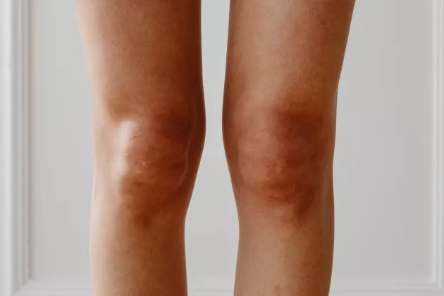
Osteoarthritis is commonly affecting the knee joint. At a late stage of the disease, the articular cartilage that covers the bone surfaces in the joint breaks down and disappears. Other tissues are also affected, e.g., the meniscus that is important for distributing load and reduce pressure on the articular cartilage. Degenerative changes of the tissue structure are starting at a microscopic level with changes in the molecular composition and the collagen structure.
This thesis focusses on meniscus and articular cartilage and the aim was to investigate imaging techniques to study tissues and degenerative changes related to knee osteoarthritis.
Magnetic resonance imaging (MRI) is suitable for examination of joints. Except standard imaging, that can be adjusted dependent on the tissue of interest, different material properties can also be measured. For example, we can measure so called relaxation times and map how they vary in the tissue and in the joint. These are dependent on the tissue structure and composition at a microscopic level and may therefore reflect degenerative changes related to osteoarthritis. Another MRI-based method is glycosaminoglycan Chemical Exchange Saturation Transfer (gagCEST) that has the potential to directly detect and measure the levels of glycosaminoglycan (GAG) that has a key role in the pressure resistance of articular cartilage.
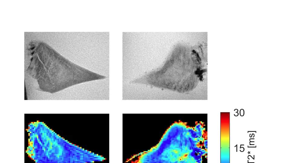
To be able to directly study the tissue structure in detail, we need microscopy techniques. A method that has not before been used for the meniscus is synchrotron radiation based microtomography. In a synchrotron facility, electrons are accelerated nearly to the speed of light to produce X-rays that can be used for example for imaging of tissue samples. Such images have very high resolution, enough to directly visualize the collagen fibers of the meniscus.
Imaging techniques are important tools for future studies of knee osteoarthritis and the role of different joint tissues.
If you want to know more, you'll find this doctoral thesis by Emma Einarsson at University of Lund's research portal.